- Cutaneous Innervation and Vasculature of the Face
- Muscles, Innervation and Blood supply of the Face
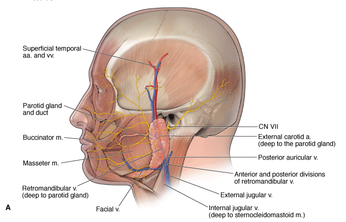
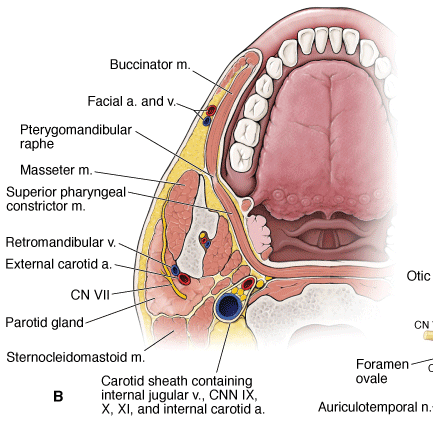
- Parotid
Vessels of the Face
The face has a very rich blood supply provided primarily from the following vessels (Figure 20-1C):
- Facial artery. Branches off the external carotid artery and courses deep to the submandibular gland; winds around the inferior border of the mandible, anterior to the masseter muscle, to enter the face. As it ascends in the face, the facial artery supplies most of the face by way of the inferior labial, superior labial, lateral nasal, and angular arteries.
- Supraorbital and supratrochlear arteries. Terminal branches of the ophthalmic artery, a branch of the internal carotid artery, which supply the anterior portion of the scalp.
- Superficial temporal artery. A terminal branch of the external carotid artery provides arterial supply to the lateral surface of the face and scalp.
- Facial vein. Formed by the union of the supraorbital and supratrochlear veins. The facial vein descends in the face and receives tributaries corresponding to the branches of the facial artery. The facial vein drains into the internal jugular vein.
 In the skull, the facial vein connects with the cavernous sinus via the superior ophthalmic vein. Unlike other systemic veins, the facial and superior ophthalmic veins lack valves, thus providing a potential pathway for the spread of infection from the face to the cavernous sinus.
Embryologic Derivation
The muscles are sometimes indistinct at their borders because they develop embryologically from a continuous sheet of musculature derived from the second branchial arch.
The muscles of facial expression arise from bones or fascia of the skull and insert into the skin, which enables a wide array of facial expression. The muscles of facial expression are located in the superficial fascia in the neck, face, and scalp. Each muscle is innervated by CN VII (except the muscles of mastication, which are innervated by CN V-3).


Muscles of the face.
Occipitofrontalis Muscle
The occipitofrontalis muscle, which lies in the scalp, extends from the superior nuchal line in the back to the skin of the eyebrows in the front. It allows for the movement of the scalp against the periosteum of the skull and also serves to raise the eyebrows.
Orbicularis Oculi Muscle
The orbicularis oculi muscle lies in the eyelids and also encircles the eyes. It helps to close the eye in the gentle movements of blinking or in more forceful movements, such as squinting. These movements help express tears and move them across the conjunctival sac to keep the cornea moist.
Orbicularis Oris Muscle
The orbicularis oris muscle encircles the opening of the mouth and helps to bring the lips together to keep the mouth closed.
Buccinator Muscle
The buccinator muscle arises from the pterygomandibular raphe in the back and courses forward in the cheek to blend into the orbicularis oris muscle in the lips. It helps to compress the cheek against the teeth and thus empties food from the vestibule of the mouth during chewing. In addition, it is used while playing musical instruments and performing other actions that require the controlled expression of air from the mouth.
Platysma Muscle
The platysma muscle extends from the skin over the mandible through the superficial fascia of the neck into the skin of the upper chest, helping to tighten this skin and also to depress the angles of the mouth. Although lying primarily in the neck, it is grouped with the muscles of facial expression.
The muscles of facial expression are innervated by the facial nerve (CN VII).
The muscles of facial expression can be organized into the following groups:
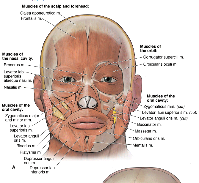
- Scalp and forehead
- Frontalis. Connects with the occipitalis muscle by a cranial aponeurosis (galea aponeurotica); and wrinkles the forehead.
- Muscles of the orbit
- Orbicularis oculi. Consists of orbital and palpebral portions, forming a sphincter muscle that closes the eyelids.
- Corrugator supercilii. Located deep to the orbicularis oculi; draws the eyebrows medially.
- Muscles of the nose
- Procerus. Wrinkles the skin over the root of the nose.
- Nasalis and levator labii superioris alaquae nasi. Flare the nostrils.
- Muscles of the mouth
- Orbicularis oris. Originates from the bones or fascia of the skull and inserts in the substance of the lips, forming an oral sphincter.
- Levator labii superioris and levator anguli oris. Raise the upper lip.
- Zygomaticus major and minor. Raise the corners of the mouth (smile).
- Risorius. Draws the corners of the lips laterally.
- Depressor labii inferioris and depressor anguli oris. Lower the bottom lip.
- Buccinator. Compresses the cheek when whistling, blowing, or sucking; holds food between the teeth during chewing.
- Neck
- Platysma. Tenses the skin of the neck and lowers the mandible. Primarily located in the neck, although it does have attachments in the lower mandible and corners of the mouth.
The corneal blink reflex is tested by touching the cornea with a piece of cotton, which should cause bilateral contraction of the orbicularis occuli muscles. The afferent limb is the nasociliary nerve of CN V-1, and the efferent limb of the reflex arch is CN VII.
Motor Nerve Supply to the Face
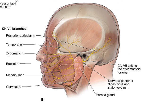
CN VII provides motor innervation to the muscles of facial expression (Figure 20-2B). The facial nerve exits the skull through the stylomastoid foramen and immediately gives off the posterior auricular nerve and other branches that supply the occipitalis, stylohyoid, and posterior digastricus muscles and the posterior auricular muscle. CN VII courses superficial to the external carotid artery and the retromandibular vein, enters the parotid gland, and divides into the following five terminal branches: temporal, zygomatic, buccal, mandibular, and cervical nerves, which in turn supply the muscles of facial expression. Other muscles of the face include muscles of mastication (temporalis, masseter, and the medial pterygoid and lateral pterygoid muscles), which are innervated by the motor division of CN V-3.
One of the most common problems involving CN VII occurs in the facial canal, just above the stylomastoid foramen. Here, an inflammatory disease of unknown etiology causes a condition known as Bell's palsy, where all of the facial muscles on one side of the face are paralyzed. Bell's palsy is characterized by facial drooping on the affected side, typified by the inability to close the eye, a sagging lower eyelid, and tearing. In addition, the patient has difficulty smiling, and saliva may dribble from the corner of the mouth. If the inflammation spreads, the chorda tympani and nerve to the stapedius muscle may be involved.
Arteries
The blood supply of the face is through branches of the facial artery

After arising from the external carotid artery in the neck, the facial artery passes deep to the submandibular gland and crosses the mandible in front of the attachment of the masseter muscle. It takes a tortuous course across the face and travels up to the medial angle of the eye, where it anastomoses with branches of the ophthalmic artery. It gives labial branches to the lips, of which the superior labial artery enters the nostril to supply the vestibule of the nose.
The occipital, posterior auricular, and superficial temporal arteries supply blood to the scalp. They all arise from the external carotid artery. The superficial temporal artery gives a branch, the transverse facial artery, which courses through the face parallel to the parotid duct.
Veins
The superficial temporal and maxillary veins join within the substance of the parotid gland to form the retromandibular vein
Download Slide (.ppt) | Print

(Figure 1–3). The facial vein joins the anterior division of the retromandibular vein to drain into the internal jugular vein. Additional details about the venous drainage pattern of the scalp and face are provided in the discussion of the veins of the neck. The facial vein communicates with both the pterygoid venous plexus and the veins in the orbit. Each of these has connections to the cavernous sinus, thus allowing infections to spread from the face into the cranium.